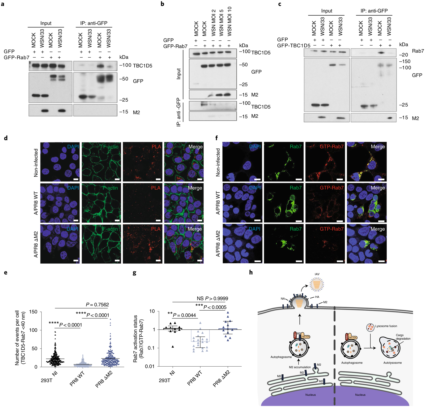
Fig. 6 |. M2 protein abrogates TBC1D5 and Rab7 interaction.

a, 293T cells were transfected with GFP or GFP-Rab7 and infected with A/WSN/33 (MOI 3) for 24 h. IP was carried out using GFP-trap resin. Inputs and IP samples were analysed by SDS–PAGE using indicated antibodies. b, 293T cells were transfected with GFP or GFP-Rab7 and infected with A/WSN/33 (MOI 2, 5 and 10) for 24 h. IP was carried out using GFP-trap resin, and inputs and IP samples were analysed by SDS–PAGE using indicated antibodies. c, 293T cells were transfected with GFP or GFP-TBC1D5 and infected with A/WSN/33 (MOI 3). At 24 h p.i. cell lysates were subjected to IP using GFP-trap resin and inputs and IP samples were analysed by SDS–PAGE using indicated antibodies. In a–c, blots are representatives from at least two independent experiments. In d and e, 293T cells were infected with A/PR8 WT or A/PR8 ΔM2 (MOI 3) and subjected to PLA staining. d, Representative images from three independent experiments show PLA signal events (red) where TBC1D5 and Rab7 proteins interact, Phalloidin (F-actin, green) and Hoechst (DNA, blue). Scale bar, 10 μm. e, Quantification of PLA signal events. Data show mean ± s.d. from one representative experiment of at least three independent experiments where at least 100 cells per condition (n = 100) were quantified. Statistical significance was calculated using one-way ANOVA with Tukey’s multiple comparisons test. f, Representative images from two independent experiments show Rab7 (green) and GTP-Rab7 (red) staining across 293T cells that express GFP-Rab7 WT and are either mock-infected or infected with A/PR8 WT or A/PR8 ΔM2 (MOI 3) for 14 h. Scale bar, 10 μm. g, Quantification of GTP-Rab7/total Rab7 ratio. Data show mean ± s.d. from one representative experiment of at least two independent experiments where at least 100 cells per condition (n = 100) were quantified. Statistical significance was calculated using one-way ANOVA with Kruskal–Wallis multiple comparisons test. h, Proposed model. IAV M2 protein abrogates interaction of TBC1D5 with Rab7, which in turn prevents fusion of autophagosomes with lysosomes. By escaping degradation at the lysosome, M2 can now assist IAV budding at the plasma membrane and support viral growth.
