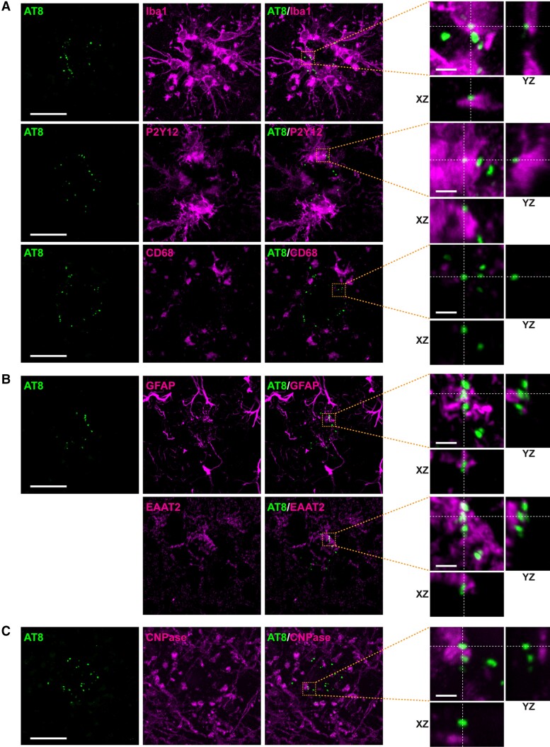
Figure 5.
Glial markers do not colocalize with p-tau 202/205/208 (AT8) in the brain of AppNLGF mice. Representative images of cortices from frozen coronal brain sections immunostained with (A) antibodies against p-tau 202/205/208 (AT8) and the microglial markers Iba1, P2Y12 and CD68 antibodies, (B) antibodies against AT8 and the astrocyte markers GFAP and EAAT2 and (C) antibodies against AT8 and the oligodendrocyte marker CNPase. The two AT8 images in B were analysed using sections co-stained with GFAP and EAAT2, with colocalizations examined in the same images as AT8 signals. Scale bars, 20 µm. Orthogonal views of different planes (yz, xz) of higher magnification images from the corresponding dashed orange squares are shown on the right; Scale bars in the orthogonal views, 2.5 µm.

