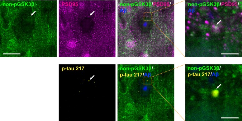
Figure 6.
Colocalization of p-tau 217, PSD95 and the active form of GSK3β in AppNLGF mice. Representative images of cortices from frozen coronal brain sections immunostained with antibodies against the active form of GSK3β (nonphospho-GSK3β) and PSD95 or p-tau 217. Aβ plaques are detected by staining with FSB. Scale bars, 20 µm. Higher magnification views of the corresponding dashed orange squares are shown in the right panel; scale bars, 2.5 µm. White arrows indicate colocalization.

