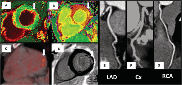
Figure 3. Focal inferolateral myocarditis with no atherosclerotic disease.

Changes (white arrows) in the native and postcontrast cardiac magnetic resonance (CMR) T1 values (A and B), 2‐deoxy‐2‐[fluorine‐18]fluoro‐D‐glucose positron emission tomography focal uptake (C), and subendocardial fibrosis on CMR late gadolinium enhancement (D). There was no significant coronary artery disease on computed tomography coronary angiography (E through G). Biochemical cardiac and inflammatory markers were low (high‐sensitivity cardiac troponin I, 2.72 ng/L; NT‐proBNP [N‐terminal pro–brain natriuretic peptide], <35 pg/mL; CRP [C‐reactive protein], 4 mg/L). Cx indicates left circumflex coronary artery; LAD, left anterior descending coronary artery; and RCA, right coronary artery.
