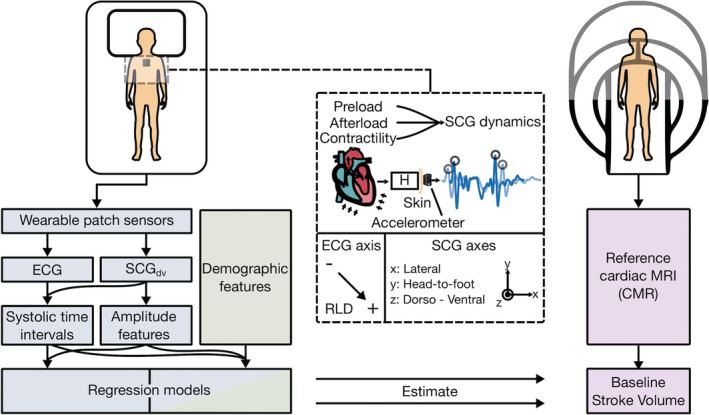
Figure 1. Concept overview.

Study design showing wearable biosensor placement when supine and asynchronous reference cardiovascular magnetic resonance imaging (CMR) measurement. Seismocardiogram (SCG) mechanistic overview detailing modulation due to cardiac physiology, acquisition with an accelerometer, and sensing axes for ECG (negative, positive, and right‐leg‐drive [RLD] electrodes), and triaxial SCG signals. Analysis pipeline, from sensor input to model estimation of stroke volume, for wearable (blue), demographic (green), and CMR (purple) data. H indicates the transfer function between the input internal sources of cardiomechanical vibration and the output SCG waveform measured on the surface of the torso; MRI, magnetic resonance imaging; and SCGdv, dorso‐ventral SCG.
