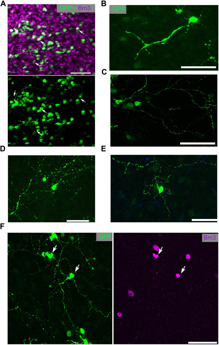
Fig. 2. IPA-stimulated MG-derived neurons display complex neuronal morphology.
(A) Retinal whole mounts stained for GFP (MG-derived cells; green) and Brn3 (purple). (B to E) Examples of the morphology of GFP+ MG-derived cells. (F) MG-derived (GFP+) cell with complex neurites colabeled with Brn3 (purple). Scale bars, 50 μm.

