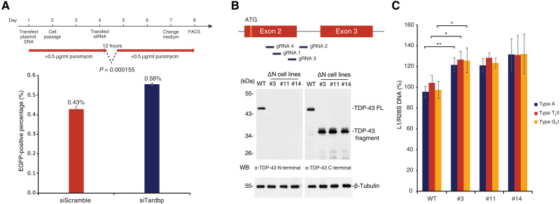
Fig. 4. TDP-43 mutation in mESCs results in increased L1 retrotransposition.
(A) mESCs treated with control and Tardbp-targeting siRNA were used for the retrotransposition assay (see Fig. 2C) and analyzed by FACS. The experimental time course is shown above. Retrotransposition frequency was increased in cells transfected with siTardbp compared with siScramble. (B) Strategy to KO TDP-43 using CRISPR-Cas9 with four gRNAs is illustrated in the top panel. The resulting clones are annotated as TDP-43 ΔN cell lines (#3, #11, and #14). These three monocloned lines were isolated, and N-terminal truncated TDP-43 was detected by WB using an anti–TDP-43 C-terminal antibody. Also see fig. S4 (C and D). (C) qPCR using primer sets targeting each active L1 subfamily (29) was performed on wild-type and ∆N lines. The DNA amount of active L1 subfamilies was increased in TDP-43 ΔN mESCs. *P ≤ 0.05 and **P ≤ 0.01.

