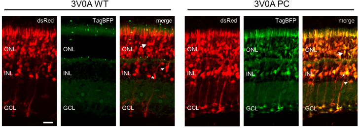
Figure 9. Nanobody expression in the murine retina representative Images of murine retina co-electroporated with CAG-dsRed and either wild-type or mutant CAG-3V0A-TagBFP plasmid.
Retinas were electroporated on postnatal day 2 and harvested on postnatal day 12. Multiple cell types show expression following electroporation: photoreceptors (arrow), bipolar interneurons (triangle), and Mueller glia (asterisk) are noted on merged images. Scale bar is 20 µm.

