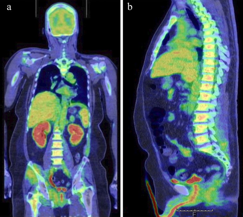
Figure 1.

Coronal (a) and sagittal (b) 18F-fluorodeoxyglucose positron emission tomography/computed tomography images showing a diffuse 18F-fluorodeoxyglucose uptake in the bone marrow, hepatosplenomegaly, and an increased 18F-fluorodeoxyglucose uptake in the spleen, both kidneys, and several muscles.
