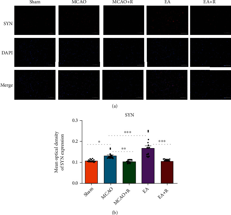
Figure 3.

IF analysis of the effects of EA on SYN expression in the contralateral cerebral cortex. (a) IF analysis showed the expression of SYN+ in each group (n = 5). (b) The mean optical density of SYN in each group (∗p < 0.05, ∗∗p < 0.01, ∗∗∗p < 0.001, SYN (red) and DAPI (blue); scale bar = 100 μm).
