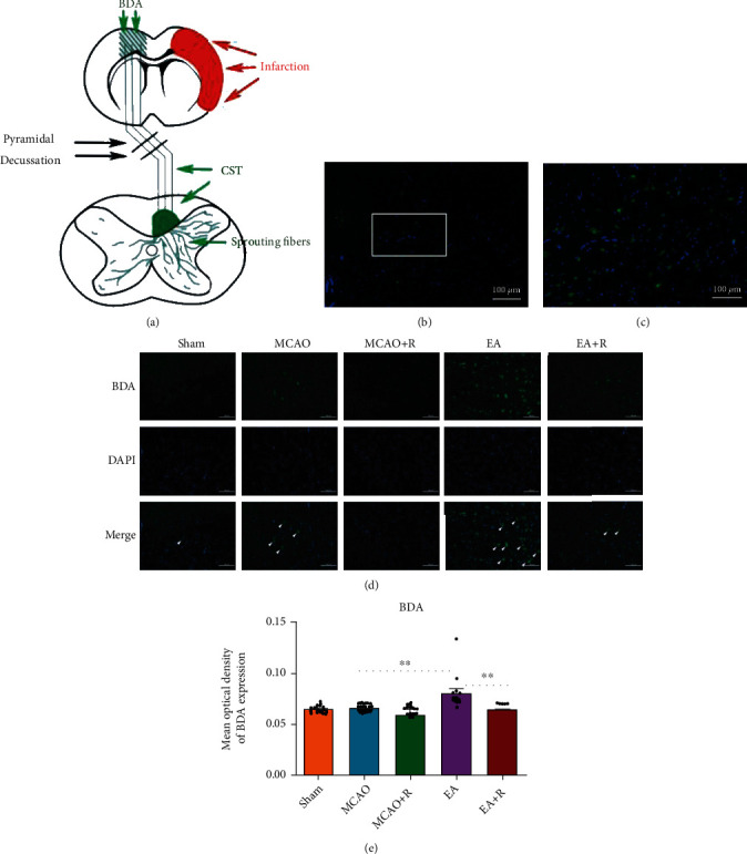
Figure 7.

Effect of EA intervention on CST axon regeneration. (a) Schematic diagram of BDA cis-neural tracer staining. (b) BDA labeling in the anterior horn region of the spinal cord gray matter in the EA group. (c) This image is a partial zoom of the white box in (b). (d) Expression of BDA-positive cells in the cervical medullary gray matter in each group (n = 5). (e) Statistical analysis of BDA-positive cells in the cervical medullary gray matter of each group (∗p < 0.05, ∗∗p < 0.01, ∗∗∗p < 0.001, arrows indicate positive cells, BDA (green), DAPI (blue); scale bar = 100 μm).
