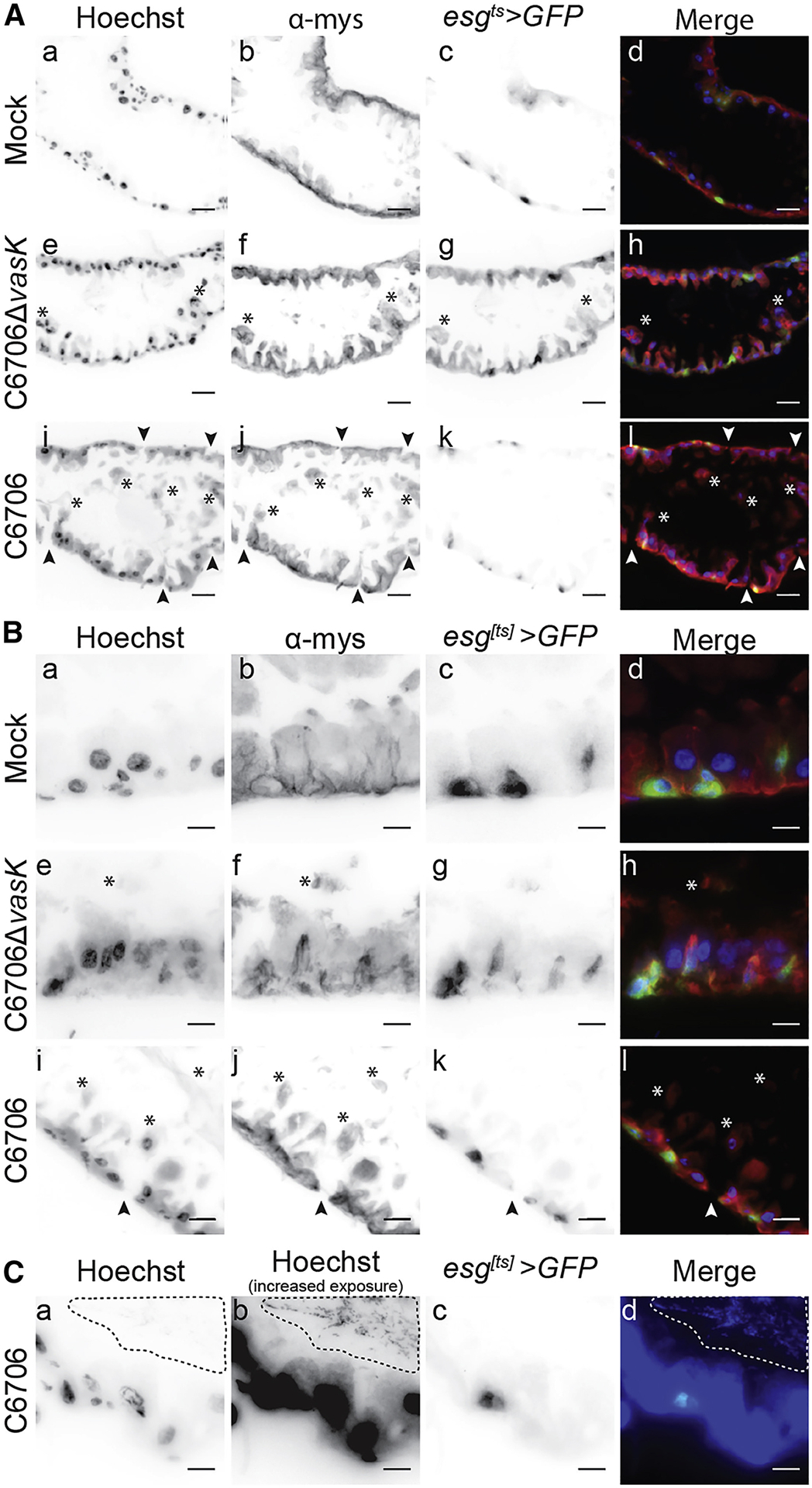
Figure 2. Disrupted intestinal homeostasis in response to the T6SS.

(A-C) Immunofluorescence of sagittal sections prepared from the posterior midgut of esgts>GFP flies mock infected or infected with C6706ΔvasK, or C6706. Hoechst marks DNA (blue), GFP marks IPCs (green), and α-mys marks the β-integrin, myospheroid (mys, red). Arrowheads indicate damage to the intestinal epithelium and asterisks denote cellular matter in the lumen. (C) Visualization of intestinal bacteria via increased exposure of Hoechst stain. The dotted line circles bacteria in the lumen. Scale bars are (A) 25μm and (B & C) 10μm.
