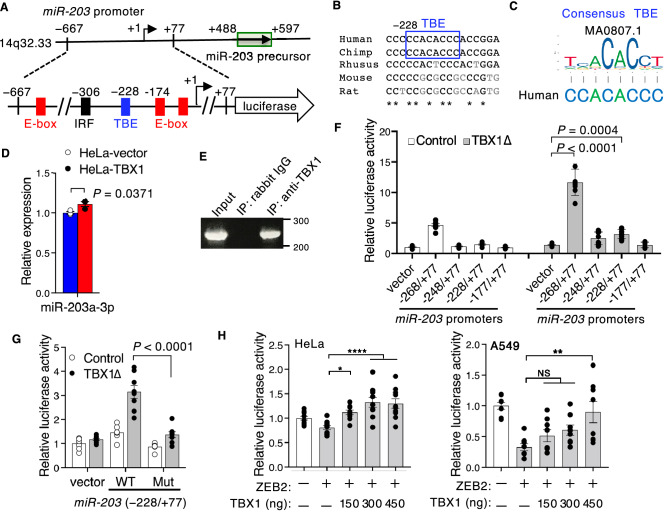
Figure 4.
TBX1 induces miR-203. (A) Schematic of the upstream promoter region of the hsa-mir-203 stem-loop on chromosome 14q32.33 and the luciferase construct of the miR-203 promoter with the location of the predicted TBE (blue box), as well as ZEB1-binding E-boxes (red boxes)24 and an IRF1-binding site (black box)50. (B) Nucleotide conservation of the miR-203 promoter, including TBE. Asterisks indicate evolutionarily conserved nucleotides in all five species. (C) Sequence of TBE from the human miR-203 promoter and the JASPAR sequence logos of TBE. (D) Expression of miR-203a-3p examined using qPCR in control HeLa‑vector cells and HeLa‑TBX1 cells. The graph shows the relative miRNA quantities (n = 3, *P ≤ 0.05; unpaired two-tailed Student’s t-test). (E) Binding of TBX1 to the miR-203 promoter (− 298/ − 66) as detected by ChIP assay using HeLa cells. Chromatin was immunoprecipitated with normal rabbit IgG or anti-TBX1 antibodies. Input control is also shown. (F) Relative luciferase activity of miR-203 promoter reporters in HeLa cells (n = 9, *P ≤ 0.05, ***P ≤ 0.001, ****P ≤ 0.0001; one-way ANOVA). (G) Relative luciferase activity of miR-203 promoter constructs (− 228/ + 77) for wild-type (WT) or mutant (Mut) TBE transiently transfected with TBX1Δ (n = 8, two-way ANOVA). (H) Relative luciferase activity of HeLa or A549 cells transiently co-transfected with the miR-203 promoter construct (− 667/ + 77), ZEB2 (150 ng), and the indicated amount of TBX1 expression vector (*P ≤ 0.05, **P ≤ 0.01, ****P ≤ 0.0001; NS, not significant; one-way ANOVA).

