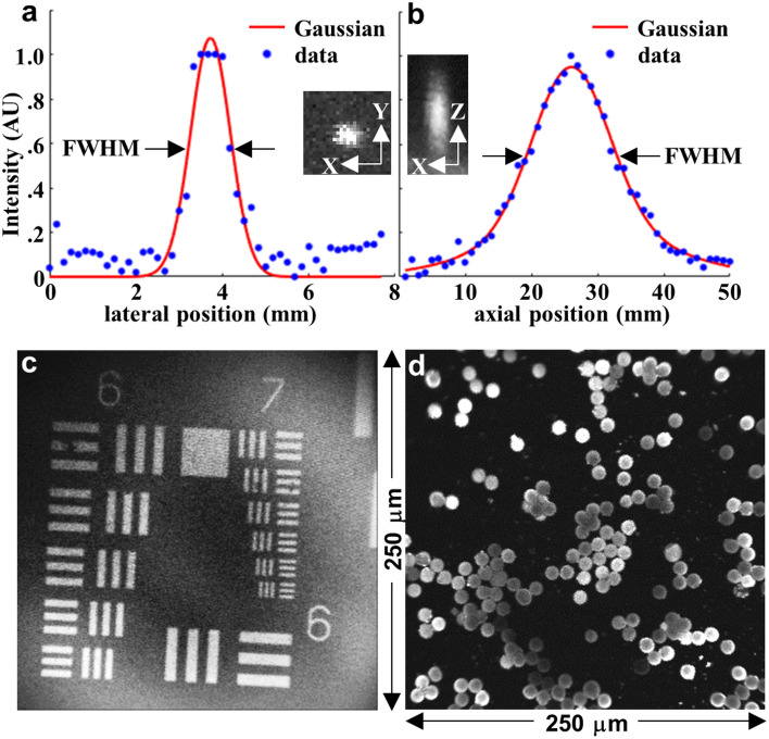
Figure 2.
Image parameters. (a) Lateral and (b) axial resolution of focusing optics was characterized by a point spread function (PSF) measured using 0.1 μm diameter fluorescent microspheres. A full-width-half-maximum (FWHM) of 1.1 and 13.6 μm, respectively, was measured. Inset: expanded view of single microspheres in the lateral (XY) and axial (XZ) directions are shown. (c) A fluorescence image collected from a standard (USAF 1951) target bars (red oval) shows that group 7–6 can be clearly resolved. (d) An image of dispersed 10 μm diameter fluorescent microspheres demonstrates an image FOV of 250 μm × 250 μm. PSFs in (a, b) were plotted using MATLAB R2019a (https://www.mathworks.com/). (c, d) Fluorescence images were collected using LabVIEW 2021 (https://www.ni.com/).

