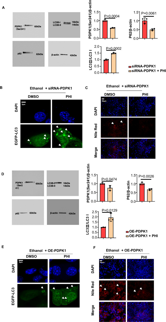
Fig. 5. Phillyrin attenuated steatosis of hepatocytes by promoting autophagy by binding with PDPK1.
A Immunoblot of cell lysates. Quantification of ratio of PDPK1 (Ser241)/β-actin, LC3II/LC3I and p62/β-actin (n = 3). B CLSM images of LC3 in AML-12 cells transfected with EGFP-LC3 plasmid (n = 3). Scale bar, 5 μM. C Nile red staining of lipid in AML-12 cells (n = 3). Scale bar, 50 μM, magnification, 400×. D Immunoblot of cell lysates. Quantification of the ratio of PDPK1 (Ser241)/β-actin, LC3II/LC3I and p62/β-actin (n = 3). E CLSM images of LC3 in AML-12 cells transfected with EGFP-LC3 plasmid (n = 3). Scale bar, 5 μM. F Nile red staining of lipid in AML-12 cells (n = 3). Scale bar, 50 μM, magnification, 400×. Data represent the mean ± SEM of three biological replicates per condition. Each dot represents a sample. P < 0 .05 was considered statistically significant. NS indicates no significance (A and D, two-tailed t-tests).

