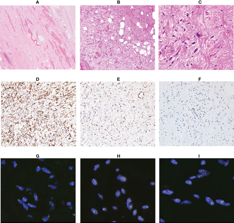
Figure 4.
Analysis of the resected specimen. The resected specimen showed an ill-defined border (A, ×100) and a myxoid and fibrous lesion with spindle cells; some spindle cells had cytological atypia (B, ×100 C, ×200). Immunohistochemical results for CD34 (D), S100 (E) and RB1 (F) (×200). Fluorescence in situ hybridization indicated no amplification of MDM2 (G) and no rearrangement of FUS (H) or EWSR1 (I).

