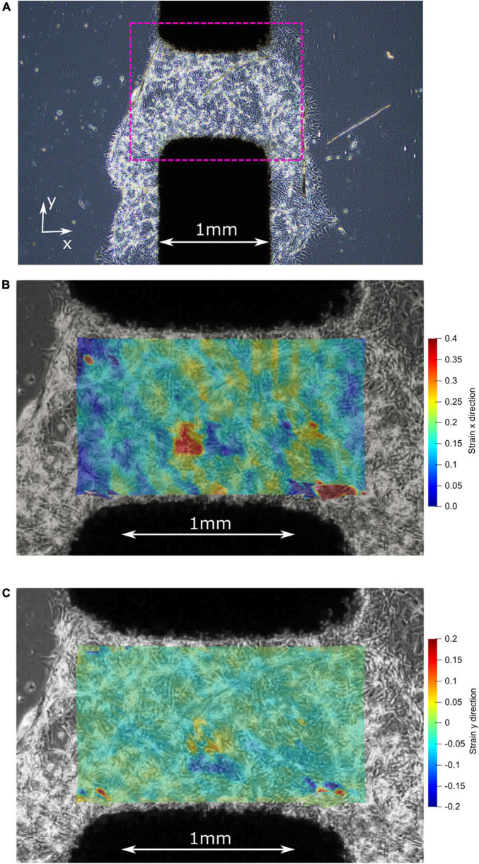
FIGURE 7.
(A) Before stretching: center of the active zone on the CID with plated 3D matrigel and pericytes. (B) After stretching: strain map illustrating the high strain (up to 40%) experienced by the pericyte cells in the x-direction. (C) After stretching: strain map illustrating the low strain (mostly 0%) experienced by the pericyte cells in the y-direction, which is the direction of the initial pre-stretch during CID fabrication.

