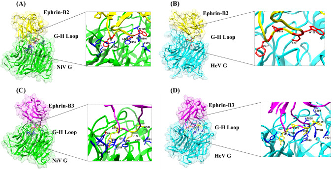
Figure 1.
Structures of (A) NiV glycoprotein/Ephrin-B2 (B) HeV glycoprotein/Ephrin-B2 and (C) NiV glycoprotein/Ephrin-B3 (D) HeV glycoprotein/Ephrin-B3 complexes with their respective interface regions. The interface residues are depicted in the square box. The NiV glycoprotein is colored in green in both the complexes and the HeV glycoprotein is colored in cyan in both the complexes. EFNB2 is colored in yellow and EFNB3 is colored in magenta.

