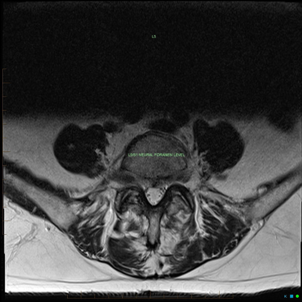
Figure 3.
Axial T2 (non-contrast) sequence further depicting a mild posterior disc bulge at the L5/S1 level without significant neural compression, which would not be sufficient to explain the degree of enhancement of the right L5 nerve root depicted in figures 1 and 4.

