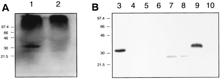
FIG. 1.
Western blot-based detection of ZnuA in culture supernatant fluid and subcellular fractions of H. ducreyi. The H. ducreyi ZnuA-reactive MAb 3F1 was used as the primary antibody. (A) Concentrated culture supernatant fluid from wild-type 35000 (lane 1) and the znuA mutant 35000.901 (lane 2). (B) Subcellular fractions from wild-type 35000 (lanes 3, 5, 7, and 9) and the znuA mutant 35000.901 (lanes 4, 6, 8, and 10). Lanes 3 and 4, whole-cell lysate; lanes 5 and 6, total cell envelopes; lanes 7 and 8, Sarkosyl-insoluble proteins from cell envelopes; lanes 9 and 10, periplasmic fraction. The aberrant migration of the ZnuA protein in the periplasmic fraction in lane 9 was caused by the presence of a high concentration of sucrose derived from the preparation of the periplasmic fraction; when this sample was diluted 1:4 in PBS, the ZnuA protein migrated at the same rate as the ZnuA protein seen in lane 3.

