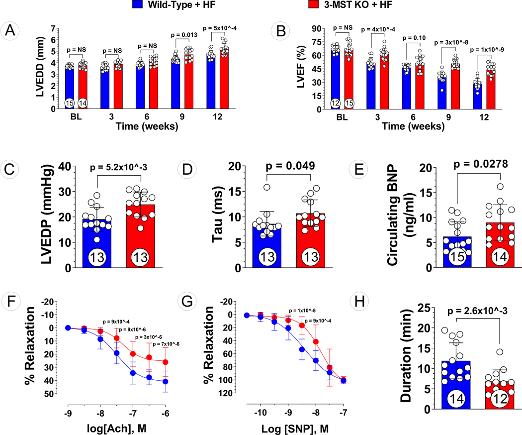
Figure 3: Exacerbated Pressure Overload HF in 3-MST KO Mice.
(A) LV end-diastolic diameter (LVEDD) and (B) LV ejection fraction (LVEF) throughout the 12 weeks study for 3-MST KO and wildtype (WT) control mice. (C) LV end-diastolic pressure (LVEDP), (D) LV relaxation constant Tau, (E) circulating B-type natriuretic peptide (BNP), (F) aortic vascular reactivity to acetylcholine (Ach), (G) aortic vascular reactivity to sodium nitroprusside (SNP), and (H) treadmill running duration in 3-MST KO and control mice at 12 weeks post TAC. Circles inside bars indicates samples size. Data in (A), (B), (F) and (G) were analyzed with ordinary 2-way ANOVA; data in other panels were analyzed with student unpaired 2-tailed t test. Data are presented as mean ± SD.

