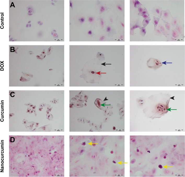
Fig. 4 .
A light microscope photomicrograph of H&E-stained cytological smears from different treated SCC4 groups. After 24 h incubation with DOX, the squamous prickle-shaped cells in (A) become swollen, demonstrating ballooning degeneration (black arrow) with disproportionated shrunken (red one) karyolitic nuclei (blue arrow) in (B). C The equivalent ghost-like (black arrowheads) cytological deteriorations retrieved from curcumin treatment confirms the necrotic cell death effect exerted by the acetone. Green arrows point out the distinctive nuclear karyorrhexis. D Meanwhile, the enhanced solubility of nanocurcumin induces apoptotic cell death, resulting in small-sized, deeply stained apoptotic bodies (yellow arrows) with pyknotic nuclei and surface blebs. Scale bar 100 µm equals × 200 magnification, while 50 µm is equivalent to × 400

