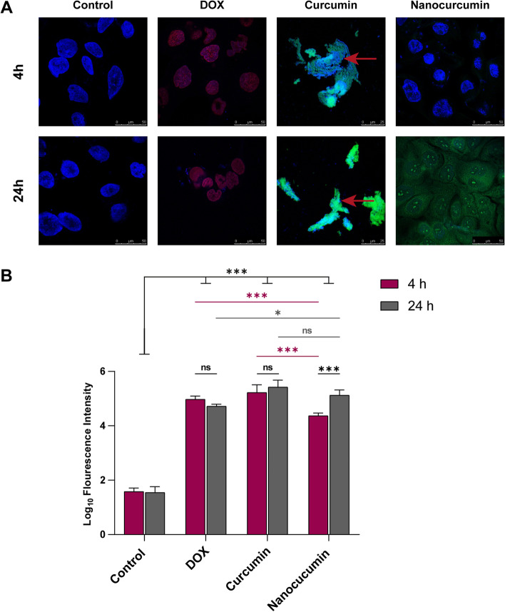
Fig. 7 .
Evaluation of the curcumin fluorescent intensity in its bulk and nano forms with cellular alterations determination, taking the DOX luminescence as a positive control. A The confocal scanning microscopy images reveal the nuclear and cytoplasmic localization of the nanocurcumin versus the nonspecific uptake of the curcumin macroparticles, whereas the DOX shows nuclear differential uptake. B The histomorphometric analyses of the fluorescence intensities, where the luminescent nanocurcumin shows a gradual increase*** in its uptake between 4 and 24 h. Meanwhile, DOX and curcumin reveal intense fluorescent signals regardless of the incubation time with drastic cytological deterioration of the necrotic matted SCC4 (red arrows in A). The data are the mean ± SD of triplicate per each time interval. Asterisk of * denotes p < 0.05 and ** implies p < 0.05, while ns of p > 0.05 points out not significance

