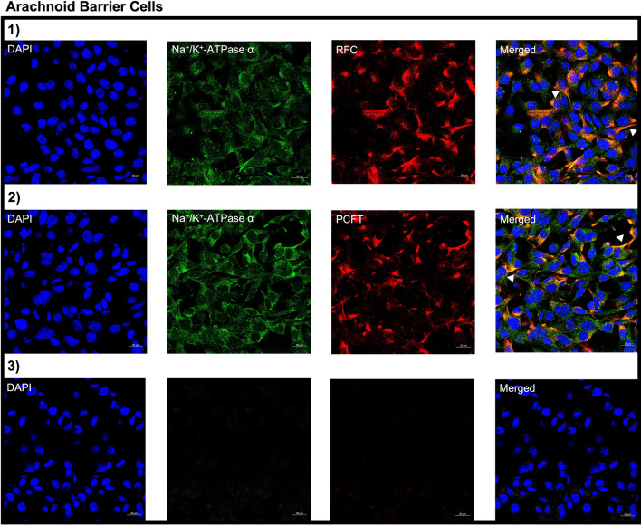
Fig. 2.
Cellular localization of RFC and PCFT in immortalized cell cultures of mouse AB. Cells were stained with the following: DAPI nuclear marker, AE390 anti-RFC (1:50) (Panel 1) or anti-PCFT (1:50) (Panel 2). To visualize the plasma membrane, cells were stained with the membrane marker Na+/K+-ATPase α (1:50). No primary antibody was used as a negative control (Panel 3). Arrows denote localization of RFC or PCFT with Na+/K+-ATPase α. Cells were visualized using confocal microscopy (LSM 700; Carl Zeiss) operated with ZEN software using an oil-immersion 63× lens

