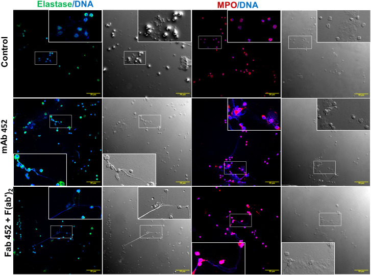
Figure 6.
Myeloperoxidase and Neutrophil elastase are associated with extracellular DNA released after CD13 crosslinking. Confocal microscopy images of human neutrophils stimulated for 240 min with mAb 452, or Fab 452 plus F(ab’)2 fragments of anti-mouse Ig, or left unstimulated (control). Cells were fixed and stained to visualize DNA (blue), myeloperoxidase (red) or neutrophil elastase (green). Each immunofluorescence image is shown together with its respective bright field micrograph. Inserts show an enlargement of a selected area of each image. Bars: 50 µm.

