
FIGURE 6.
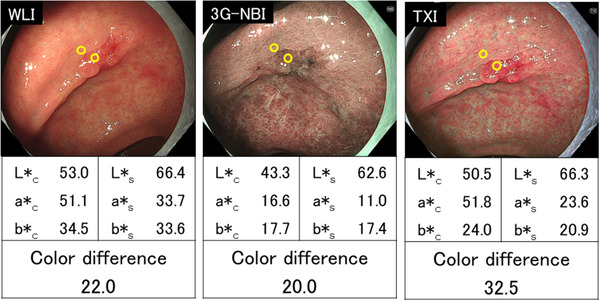
A representative case in which the color difference was larger in texture and color enhancement imaging (TXI) and smaller in third‐generation narrow band imaging (3G‐NBI) than in white light imaging (WLI). A depressed lesion (0–IIc) in the lower third of the stomach (25 mm in diameter) was observed. The final histopathological diagnosis was well‐differentiated adenocarcinoma, confined to the mucosa. The color difference was 22.0 in WLI, 20.0 in 3G‐NBI, and 32.5 in TXI