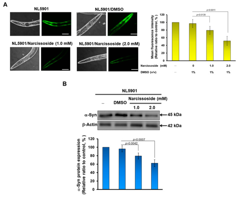
Figure 11.
α-Synuclein accumulation in nematode muscle cells is diminished by narcissoside (NCS)-induced autophagy. (A) NL5901 nematodes at the L3 stage were treated with NCS for three days. The fluorescence intensity exhibited by accumulating α-synuclein in muscle cells was then analyzed using a fluorescence microscope (Leaf panel). The fluorescence intensity was quantified using ImageJ software (Right panel). (B) The expression of α-synuclein in each group of nematodes in (A) was analyzed by Western blotting. β-actin is an internal loading control (top panel). The fluorescence intensity was quantified using ImageJ software (bottom panel). (C) DA2123 nematodes at the L3 stage were treated with NCS for three days. The dots formed by the autophagy marker LGG-GFP in seam cells were observed (Leaf panel) and counted by a fluorescence microscope. The right panel is the average result of the fluorescent dot count analysis of all seam cells in each group of worms.


