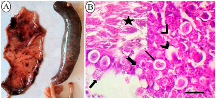
Figure 1.
The pathological findings in ceca of E. tenella naturally infected broiler chickens. (A): Gross appearance of ceca showing severe congestion and dilatation and filled with bloody contents with friable cecal wall and loss of its lining mucosa. (B): Histopathological cecal section illustrating the distinct developmental stages of E. tenella; mature oocyst with a central nucleus and oocyst wall (Chevron arrow); immature oocyst with a central nucleus and oocyst wall (thin arrow); macrogametes with bordering eosinophilic bodies and a central nucleus (Bent-up arrow); microgametes (Thick arrow); developing schizont (notched arrow), mature second stage schizonts (star) arranged in groups and having several crescent-shaped merozoites (H&E, bar = 50 µm).

