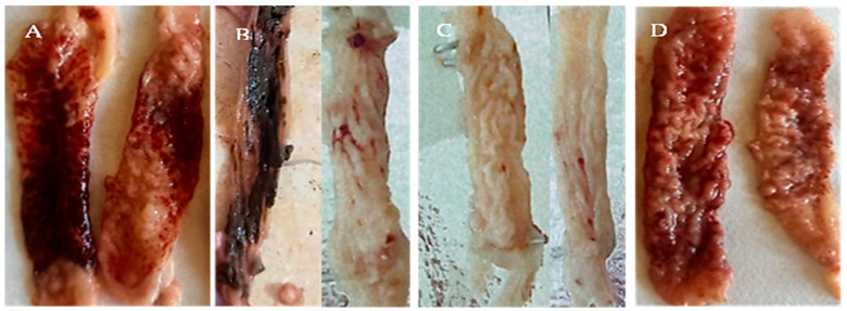
Figure 4.
Gross pathological findings in the ceca of E-infected 3-week-old coccidia-free chickens (7 days after infection) tenella oocysts sporulated in various solutions. (A) Lesion score + 4 was caused by E. tenella oocysts that sporulated in 2.5% K2Cr2O7, revealing a sheet of coagulated blood covering the mucosa, complete loss of cecal corrugations, and ulceration. (B) Lesion score +1 induced by E. tenella oocysts sporulated in allicin (90 mg/mL) solutions; slight thickening of the cecal wall; cecal contents are relatively normal. (C) Lesion score +2 was induced by E. tenella oocysts that sporulated in an alcoholic garlic extract (180 mg/mL) solution, revealing a moderately thick cecal wall, scattered patechae on the mucosal surface, and tenacious cecal contents. (D) Lesion score +3 induced by E. tenella oocysts which sporulated in KOH 5%; ceca had a markedly thickened wall, eroded mucosal surface, and bloody cecal contents.

