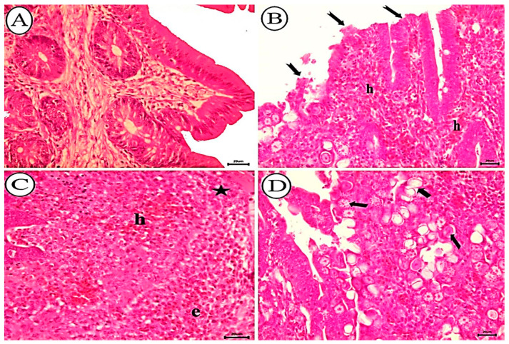
Figure 6.
Photomicrograph showing histopathological characteristics of cecal tissues. (A) Negative control group of non-infected birds showing normal tissue appearance. (B–D) Birds infected with non-treated oocysts. (B) Significant epithelial shedding (notched arrow) and diffuse mucosal bleeding (h). (C) Diffuse mucosal hemorrhages (h), eosinophils (e), and mononuclear cell infiltration (star). (D) Various coccidial stages in the villar epithelium and lamina propria (arrow), H&E stain, scale bar = 20 µm.

