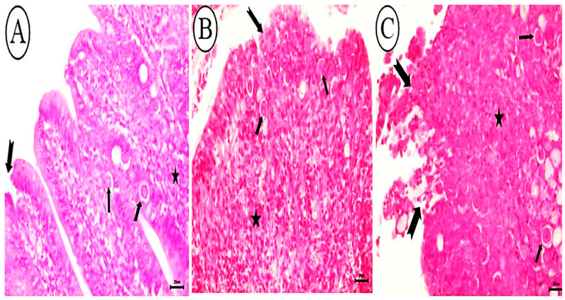
Figure 7.
Photomicrograph depicting histopathological characteristics of cecal tissues. (A). Birds infected with allicin-treated oocysts exhibited a significant reduction in the number of coccidial stages (arrow), accompanied by slight epithelial necrosis at villar tips (notched arrow) and infiltration of mononuclear cells (star). (B) Birds infected with garlic extract-treated oocysts display reduced epithelial necrosis (notched arrow), coccidial stages (arrow), and mononuclear cells in the lamina propria (star). (C) Birds infected with KOH-treated oocysts exhibiting villar necrosis (notched arrow), mononuclear cells (star), and parasitic stages in the lamina propria (arrow), H&E stain, bar = 20 µm.

