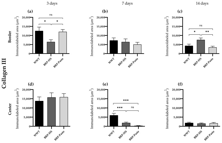
Figure 7.
Quantification of collagen type III by immunolabed area (μm2) at border (a–c) and central (d–f) regions (dermis) of the wounds in each experimental group for the periods of 3 (a,d), 7 (b,e) and 14 (c,f) days of treatment. Statistical difference compared to the WWT group was obtained by one-way ANOVA followed by Newman-Keuls test in which * p < 0.05, ** p < 0.01, and *** p < 0.001 (n = 6). ns = no significance.

