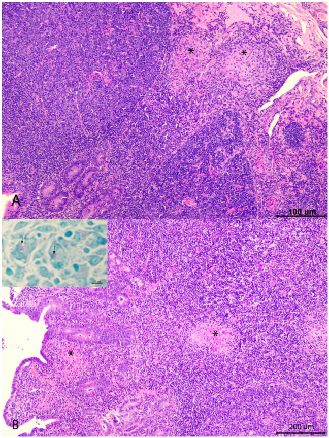
Figure 2.
Granulomatous lesions associated with paratuberculosis infection found in the necropsied goats. (A): focal lesion, formed by small granulomas (*) located exclusively in the interfollicular area of the Peyer’s patches. Hematoxylin-eosin (HE) (B): multifocal lesion, composed of granulomas (*) located both in the interfollicular area on the Peyer’s patches and related lamina propria. HE. Inset: small numbers of acid-fast bacilli (arrows) seen in the lesion located in the lamina propria. Ziehl–Neelsen stain (inset).

