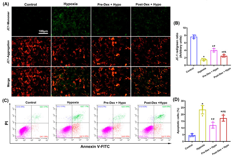
Figure 5.
The effect of Dex on mitochondrial function and apoptosis in cultured hippocampal neurons following hypoxia. (A) Representative images of the MMP in hippocampal neurons using the mitochondrial probe JC-1. (B) The MMP is represented by the ratio of red to green JC-1 fluorescence. (C,D) Apoptosis was detected and analyzed using flow cytometry by staining cells with FITC-Annexin V/PI. Results are expressed as mean ± SD (n = 4 in each group). Each data point in the bar graph is also presented. * p < 0.05 versus the Control group; # p < 0.05 versus the Hypoxia group; & p < 0.05 versus the Pre-Dex group.

