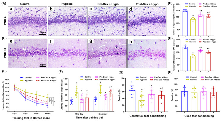
Figure 6.
The effect of Dex on histopathological changes and cognitive function in the hippocampus following neonatal hypoxia. (A,C) Nissl staining was performed to evaluate hippocampal neuronal injury and morphological changes at 1 d (PND4) and 28 d (PND31) after neonatal hypoxia. (B,D) Quantification of the number of Nissl-positive cells present in the CA1 region of the hippocampus. (E) Performance with the Barnes maze test in training sessions. (F) Performance with the Barnes maze test in the memory phase. (G) Contextual fear conditioning test. (H) Cued fear conditioning test. Results are expressed as mean ± SD (n = 4 in each group for Nissl staining data; n = 12 in each group for Barnes maze and fear conditioning test data). Each animal data point in the bar graph is also presented. * p < 0.05 versus the Control group; # p < 0.05 versus the Hypoxia group.

