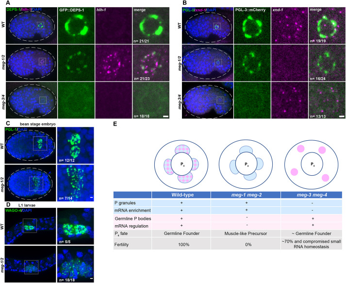
Fig. 7.
Primordial germ cells exhibit somatic-like characteristics in meg-1 meg-2 mutants. (A) Photomicrographs of bean-stage embryos of the indicated genotypes expressing DEPS-1::GFP and probed for hlh-1 RNA. Inset depicts a primordial germ cell. Embryos were scored from one independent experiment in which mutant and control animals were processed in parallel. All wild-type (21/21) and meg-3 meg-4 (18/18) bean-to-comma-stage embryos did not express hlh-1, while 21/23 meg-1 meg-2 did express hlh-1. (B) Photomicrographs of bean-stage embryos of the indicated genotypes expressing PGL-3::mCherry and probed for xnd-1 RNA (which is transcribed in PGCs at this stage). Inset depicts a primordial germ cell. Embryos were scored from two independent experiments for meg-1 meg-2 and one experiment for meg-3 meg-4 in which mutant and control animals were processed in parallel. All wild-type (19/19) and meg-3 meg-4 (13/13) bean-stage embryos expressed xnd-1, while 16/24 meg-1 meg-2 embryos did not express xnd-1. (C) Maximum projections of bean-stage embryos of the indicated genotypes stained for PGL-1. Inset shows the primordial germ cells. Embryos were scored from one experiment in which mutant and control animals were processed in parallel. All wild-type embryos (12/12) had two PGL-1-positive cells and 7/14 meg-1 meg-2 embryos had more than two PGL-1-positive cells. Dashed white line indicates embryo boundary. Dashed square indicates P4 descendants. (D) Maximum projections of germ cells from unfed L1 larvae expressing the germ granule marker 3×FLAG::GFP::WAGO-4. Embryos were scored from one experiment in which mutant and control animals were processed in parallel. All wild-type embryos (5/5) had two WAGO-4-positive cells and all meg-1 meg-2 embryos (18/18) had more than two WAGO-4-positive cells. (E) Working model: schematic and table summarizing P4 phenotypes based on this study and on Wang et al. (2014) and Ouyang et al. (2019). P granules are depicted in blue, germline P-body in pink and their merge in a checkered pattern. Note that P granule and germline P-body proteins also exist in a more dilute state in the cytoplasm. See text for additional details. Scale bars: 1 µm.

