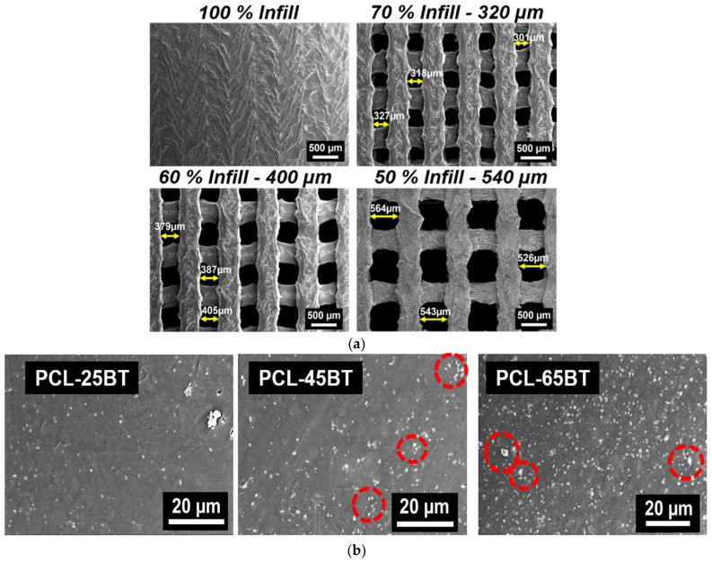
Figure 7.
SEM analysis of the scaffolds using secondary electron detector. (a) SEM of the non-porous and porous 3D-printed PCL-45BT scaffolds. The pore sizes of the scaffolds were varied by changing the infill percentage in the slicing software. (b) SEM of various PCL-BT filament cross-sections showing the uniform dispersion of the white BaTiO3 particles in the PCL matrix.

