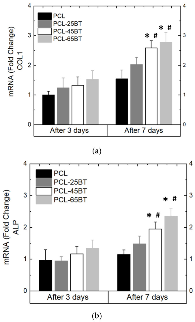
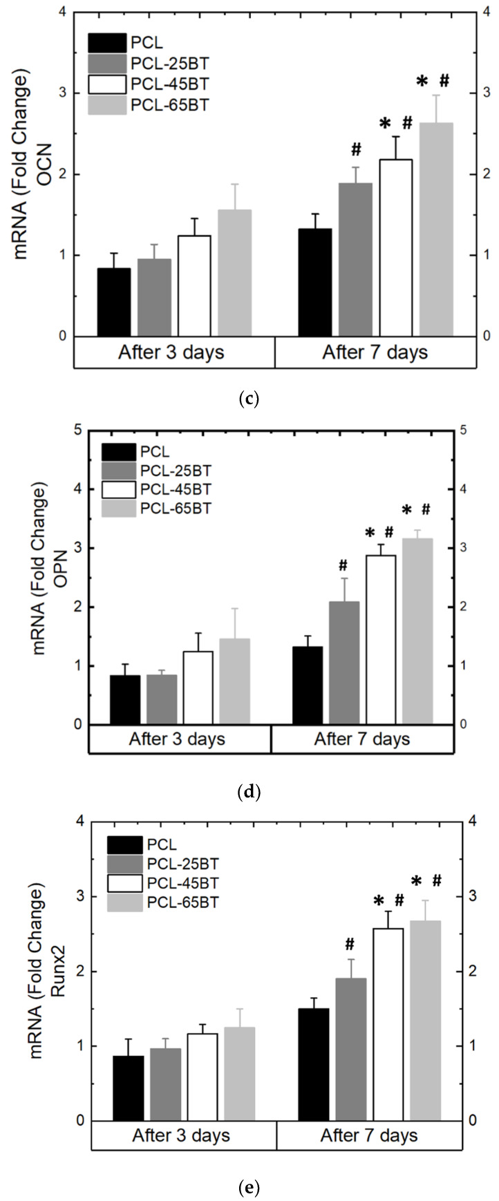
Figure 13.
Differentiation properties of MC3T3 cells as cultured on the piezoelectric scaffolds. The following osteogenic gene markers were quantified: (a) Col 1 (b) ALP (c) OCN, (d) OPN, and (e) Runx 2 and expressed as fold change with respect to the control; 3 MHz US was used per the routine for all the differentiation assays. * means statistically significant (p < 0.05) with respect to the control (PCL) in the same group. # means statistically significant (p < 0.05) with respect to the fold changes of the same specimen in the “after 3 days” group.


