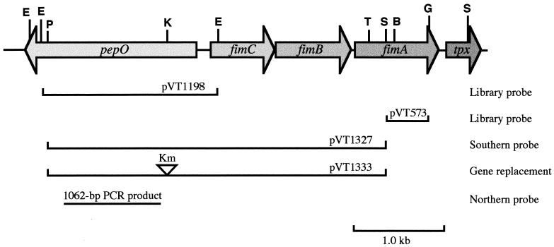
FIG. 1.
Physical map of pepO and the fimA operon region of S. parasanguis. The diagram shows a simplified restriction map of a 5.1-kb DNA fragment containing pepO, three genes (fimC, fimB, and fimC) of the fimA operon, and tpx. Arrows indicate the locations and orientations of the ORFs. The lines below the map indicate the sizes and locations of probes and plasmid inserts. The Kmr marker, shown by an open arrowhead, was inserted at the KpnI restriction site of pepO. Restriction sites were as follows: B, BamHI; E, EcoRI; G, BglII; K, KpnI; P, PstI; S, SalI; and T, SstI.

