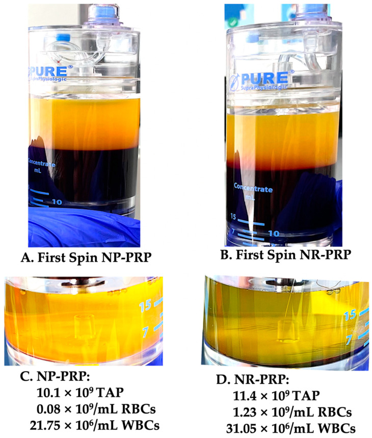
Figure 3.
Visual aspects after the First and Second spin of NP-PRP and NR-PRP Preparations. After the first centrifugation spin, no differences occur for NP-PRP (A) and NR-PRP first phase preparation (B). During this cycle, the unit of whole blood is sequestered in a platelet poor plasma (PPP) fraction, thin buffy coat layer, and a packed RBC layer. After the second spin, the plasma in the second device chamber is removed until the remaining volume in each device is in accordance with the total calculated treatment volumes for both bioformulations. Thereafter, the cells at the bottom of the chamber are resuspended with the remaining plasma. Laboratory data present the differences between the NP-PRP (C) and NR-PRP (D) fractions. Note, the broader RBC layer in the NR-PRP vial (D) when compared to the NP-PRP preparation (C). Furthermore, the NR-PRP buffy coat stratum on top of the RBCs is more noticeable (PurePRP-SP® device, used with permission from EmCyte Corporation, Fort Myers FL, USA) (Abbreviation: TAP: total available platelets).

