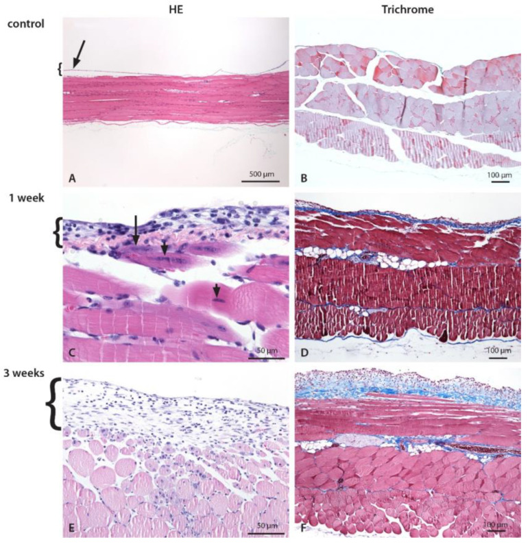
Figure 3.
Histological changes of the abdominal wall and the parietal peritoneum after CHX treatment. Inflammation with mixed cell infiltrate composed of lymphocytes, macrophages, and neutrophils. Numerous necrotic muscle cells comprising up to 25% of the depth of the muscle wall infiltrated with inflammatory cells and an increased amount of intercellular fluid (edema) were present in both groups. Edema, fibroblasts, and consequent fibrosis were more pronounced in 3-week CHX-treated mice. The abdominal wall of control (A,B), 1-week (C,D), and 3-weeks (E,F) CHX-treated mice. (A,B). Normal abdominal wall with a longitudinal section of striated muscles covered by a thin layer of the parietal peritoneum, which is partially detached (technical artifact) (arrow). (C) On the peritoneal surface, there is a mild to moderate exudate composed of neutrophils, macrophages, lymphocytes, and scarce fibroblasts. The inner layer of the striated muscle cells, with necrotic (arrow) and damaged (arrowhead) striated muscle cells, and mild edema. (D) Early fibrosis is present. (E) On the peritoneal surface, there is a mild to moderate exudate composed of neutrophils, scarce mononuclear cells (macrophages, lymphocytes), and fibroblasts. Inflammatory cells invade the edematous abdominal wall and surround necrotic striated muscle cells encompassing up to 1/4 of the abdominal wall. Necrotic muscle cells are infiltrated with inflammatory cells and an increased amount of intercellular fluid (edema). (F) Edema and fibrosis on the peritoneal surface and in the abdominal wall.

