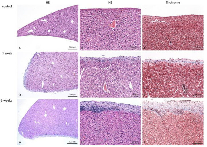
Figure 4.
Liver of control (A–C), 1-week (D–F), and 3-weeks (G–I) CHX-treated mice with hematoxylin eosin and trichrome staining. In control mice, the normal liver is seen. In the 1-week CHX-treated mice edema of liver parenchyma, diffuse moderate exudate on the surface composed of mononuclear cells and fibrosis is present. In 3-weeks CHX-treated mice edema of liver parenchyma, diffuse moderate exudate on the surface composed of mononuclear cells and more intense fibrosis are present. Legend: HE: haematoxylin eosin, (A,D,G,I) 40×, (B,C,E–H) 100×.

