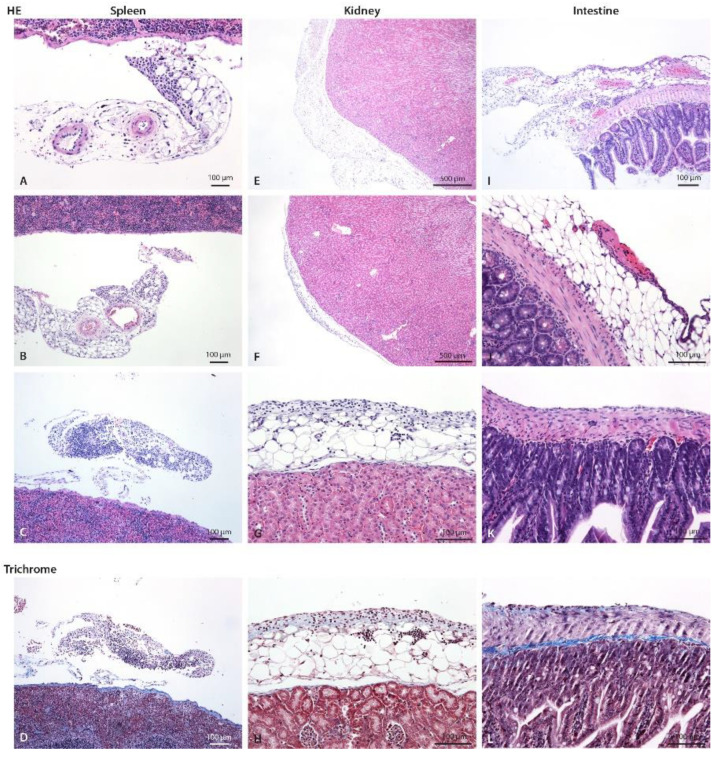
Figure 5.
Spleen, kidney, and intestine of control and CHX-treated mice with hematoxylin eosin and trichrome staining. (A) Mild focal infiltrate composed of neutrophils and scarce lymphocytes and macrophages on perisplenic fat tissue/mesenterium (arrow) in control mice, (B) in 1-week and (C) 3-weeks CHX-treated mice. (D) Early fibrosis (blue). (E) A normal perirenal fat tissue at one pole (arrow) and (F) focal infiltrate on perirenal fat (arrow) at the other pole of the kidney in 3-weeks CHX-treated mice. Renal capsule and parenchyma are normal without inflammatory cells in the fat. (G) A closer view, infiltrate is composed of neutrophils, mononuclear cells, and fibroblasts. (H) Early fibrosis (blue). A similar infiltrate was present also in 1-week CHX-treated mice. (I) In the intestinal fat (subserosa) and mesenterium, there is mild to moderate infiltrate composed of neutrophils, macrophages, lymphocytes, and scarce fibroblasts, and focal fibrinous exudate on the surface of intestinal peritoneum consistent with fibrinous purulent peritonitis (arrow) in 3-weeks CHX-treated mice. (J) A closer view: scarce neutrophils and fibrinous exudate on intestinal serosa (arrow). (K) Mixed cell exudate on the intestinal wall. (L) Early fibrosis is present.

