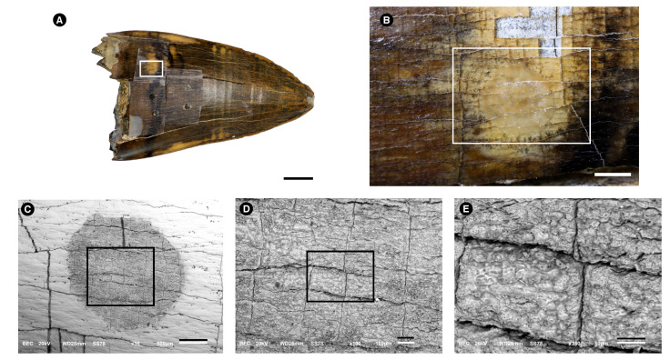
Figure 1.
Images of a Purussaurus sp.(Crocodylia, Alligatoridae) tooth used to obtain superficial enamel acid etch samples. Images were made with photograph cameras (A,B) and with a Scanning Electron Microscope using Back-scattered-electrons (BSE) mode (C–E). Rectangles are always amplified in the next figure of the series. (A). Purussaurus sp. (Crocodylia, Alligatoridae) tooth picture taken without magnification lens. The white arrow indicates enamel cracks. Bar = 1 cm. (B). Details of the white rectangle depicted in (A). Bar = 1 mm. In the lower area of the figure, some “squares” have fallen, showing the dentine below the enamel. (C). Amplification of the white rectangle shown in (B). The circular area (gray) was etched with the acid. Bar = 500 m. (D). Amplification of the rectangle shown in (C). Bar = 100 m. (E). Amplification of the rectangle shown in (D). Bar = 50 m.

