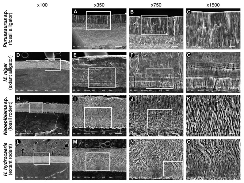
Figure 5.
Images obtained by SEM analysis of enamel cross-sections from the four (4) specimens Purussaurus sp., Melanosuchus niger, Neoepiblema sp., and Hydrochoerus hydrochaeris. (A–C), the enamel of Purussaurus sp. is formed by rod-like structures that run roughly perpendicular to the enamel surface and dentine-enamel junction (DEJ), having a width of approximately 5 m. (D–G), Melanosuchus niger has a typical aprismatic structure, showing growth lines that are likely to be the equivalent to the mammalian Retzius lines. These lines were localized near the DEJ and are likely to represent periods of physiological stress during enamel synthesis. (H–K), the enamel of Neoepiblema sp. is formed by prisms that are roughly parallel and perpendicular to the DEJ and enamel surface. The DEJ exhibits an irregular surface. (L–O), the enamel of Hydrochoerus hydrochaeris shows 2 distinct patterns of prims arrangement. The prisms run nearly parallel from the DEJ and extend approximately 25–30 m near the DEJ. A thin layer of parallel prisms is also observed near the enamel surface. In most parts of enamel, the prisms exhibit a pattern of intense decussation.

