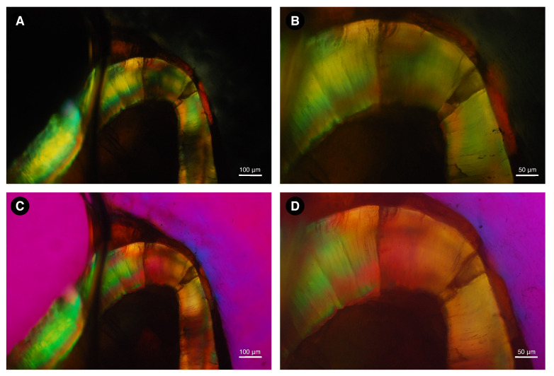
Figure 7.
Image of a ground section of dental enamel sample from Neoepiblema sp. taken under polarizing microscopy with water immersion. Without an interference filter, dental enamel shows high interference colors (A,B), indicating a relatively high ground section thickness. With the Red I filter, the addition interference color is shown at the −45° diagonal position (C), and the subtraction interference color is shown at the +45° diagonal position (D), indicating negative birefringence.

