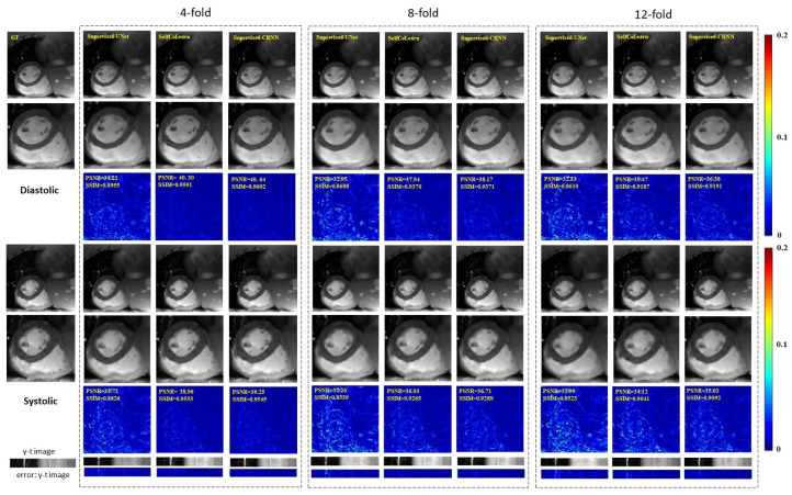
Figure 4.
Reconstruction results of different methods (Supervised U-Net, SelfCoLearn, and Supervised CRNN) at 4-fold acceleration, 8-fold acceleration, and 12-fold acceleration. The first row and fourth row show the ground truth (fully sampled image) and the reconstruction images of respective methods in the diastolic (the 10th frame of the image sequence) and systolic (the 5th frame of the image sequence), respectively. The second row and fifth row show their corresponding enlarged images in the heart regions. The third row and sixth row plot the error images of the corresponding methods. The last two rows show y-t images (the 40th slice along the dimensions of y and t) and the corresponding error images.

