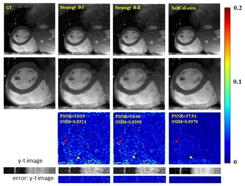
Figure 6.
Ablation studies utilizing different training strategies at 8-fold acceleration. The first row shows the ground truth (fully sampled image), and the reconstruction images of strategy B-I, strategy B-II, and proposed SelfCoLearn (10th frame). The second row shows their enlarged images in the heart regions. The third row plots the error images of respective methods. The last two rows show y-t images (the 40th slice along the dimensions of y and t) and the corresponding error images.

