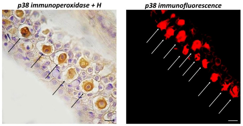
Figure 2.
Longitudinal sections (5 μm thick) of zebrafish skin, Sections are immunohistochemically treated with MAPK p38. Immunoperoxidase counterstained by H, 100×, scale bar 100 μm. Immunofluorescence, 40×, scale bar 40 μm. Immunoreactive CCs for MAPK p38 (arrows) appear evident, with a large core centrally located. They are well organized in the intermediate epidermal layer as shown by counter colouring with H.

