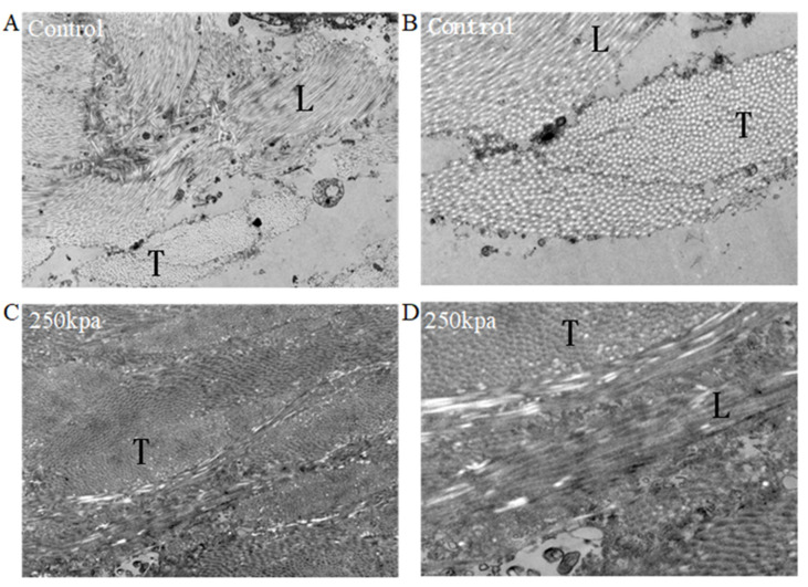
Figure 3.
Representative TEM of unfused and fused small intestine. (A,B) Electron micrograph of control (unfused) small intestine (A: magnification 3000×, B: magnification 7000×). (C,D) Electron micrograph of fused small intestine at 250 kPa ((C): magnification 3000×, (D): magnification 7000×). T, transverse-section of collagen fibrils; L, longitudinal-section of collagen fibrils.

